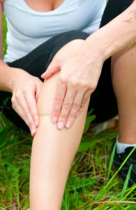
Your medial collateral ligament (MCL) is a large flat ligament band located on the inside (medial aspect) of the knee. It is one of the four ligaments that stabilise the knee joint, alongside the Lateral Collateral Ligament (LCL) and the anterior and posterior cruciate ligaments within the knee. It connects the top of the shin bone (tibia) to the bottom of the thigh-bone (femur) and works to resist forces applied to the outside of the knee (valgus forces), such as when a ball hits the outside of the leg or knee. This is also the main way it is injured.

MCL injuries occur as a result of stretching and stressing the ligament past the point that it can safely handle. Often this results from excessive force to the outside of the knee (valgus force) and various twisting motions at the legs and knees. Examples include:
Those with muscle weakness, ligament laxity or a history of knee injuries are typically at a greater risk of sustaining an MCL injury.
The different levels of severity of injury include a ligament sprain, a partial tear, or a complete rupture. Symptoms can include:
When the injury first occurs, it’s important to stop physical activity and avoid walking on the affected knee where possible. Following the PRICE principles (protection, rest, ice, compression and elevation) can help reduce the initial pain and swelling. If you have had a rupture or significant tear to your MCL, you may require surgery. It’s important that you have a diagnosis to confirm the extent of your injury, which may involve having an ultrasound, x-ray or MRI imaging. Your podiatrist can help you gradually rebuild the strength in your knee and surrounding muscles, and improve the range of motion in the joint, which will likely be restricted following your injury. This will likely focus on strengthening your quadriceps, hamstrings, calves, hips and pelvic muscles. Your podiatrist will work with you to both help your recovery and reduce the risk of re-injury in the future. This may look at the use of orthotics to control motion at the feet and legs, assessing the stability of your footwear, assessing your walking/running technique, teaching you how to strap your knee when needed, bracing, and physical therapy (strengthening and stretching).
.png)
Since introducing shockwave therapy, we’ve helped many of our patients avoid surgery for certain conditions. Here's what you
need to know about shockwave treatment and how it works.
.png)
This Mother’s Day, consider a practical, medically safe, and confidence-boosting gift: a professional KeryFlex nail restoration treatment. It’s a simple, effective, and medically safe way to instantly transform the appearance of toenails.
Keeping your family on their feet and helping them to walk, run, play and exceed their goals is why we love getting up in the morning.
Ground Floor, One Health Building
122 Remuera Rd, Remuera
Auckland 1050, New Zealand
| MON - FRI | 7:30am – 6:30pm |
| SAT | 8:30am – 4:30pm |
| SUN | Some availability |
Make an Appointment
Online Schedule